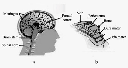
The meninges (singular meninx) are a three-part system of membranous layers or lamellae (see figure below) that form a bag-like envelope surrounding the central nervous system (brain and spinal cord), and which contain cerebrospinal fluid (CSF). Together, the cardinal function of the meninges is to protect the central nervous system (CNS) from mechanical damage and support associated vascular structures. There are subtle functional differences between the three layers. The outermost layer is the dura mater (also called the the thecal sac), which is made up of thick fibrous connective tissue. Its superficial layer serves as the inner periosteum of the skull (also referred to as the endocranium). Forming the dural sac extending from the foramen magnum (large opening in the occipital bone of the skull) to the coccyx (the ‘tailbone’), it envelopes and supports the dural (cerebral/venous) sinuses and carries blood from the brain toward the heart. The periosteum is attached by Sharpey’s perforating fibers to the skull that enter deeply into the bone (fibers discovered by William Sharpey, 1802-1880, in 1846). The pia mater forms the innermost layer, which is thin (a monolayer of cells thought to be impermeable to fluid) adhering directly to the surface of the brain and spinal cord. It is penetrated by blood vessels that travel to the ventricles of the brain and to the spinal cord. It also provides lubrication for tissues of the CNS, as well as preventing irritation of the CSF. The middle layer. with a spider-like structures (villi), is the arachnoid matter containing blood vesels that runs over the surface of the brain, but does not enter the sulci. The arachnoid villi (or granulations) reabsorb the CSF into the saggital dura lying within the dura. Together the arachnoid and the pia are called the leptomeninges, and they are separated by the subarchnoid space through which CSF flows. During embryological development, the dura mater arises from paraxial mesoderm surrounding the neural tube, and the pia mater and arachnoid mater from neural crest cells. Pathological processes associated the meninges include hemorrhage, infection and tumor formation. Also, the meninges protrude from the vertebral column in the case of spina bifida with meningcoele (spina bifida cystica). Cells of the pleuripotent neural crest migrate in embryo to give rise to various structures including the meninges of the brain and spinal cord (others are the ganglia of the dorsal roots, autonomic nervous system, and some cranial nerves, as well as the myelin sheaths of peripheral nerves).