A sensory system that detects position and motion (specifically acceleration of the head), and which is a crucial organ of balance. It is comprised of bilateral semicircular canals, the utricles and otoliths, all located in the inner ear, and includes the vestibular ganglion, and the vestibular nuclei in the brain stem as well as the medially descending vestibulospinal projections. Relative to other sensory systems, there is a dearth of knowledge about the development of the vestibulum in humans. However, like the cochlea, the system originates from the otic placode (a plate-like structure showing as a thickening of the ectoderm) in the early embryo, and thus is considered to be one the first sensory organ to develop. Both organs use the same specialized hair cells to convert physical motion into neural impulses: for the cochlea, the motion is airborne sounds and for the vestibular system it involves not only motions arising from head movements, but also inertial effects due to gravity and ground-borne vibrations. Vestibular disorders and dysfunctions increase with age, and in adults can be associated with autoimmune problems and allergies. Signs and symptoms include dizziness and vertigo, as well as nausea, headaches and motion sickness. Inevitably there are balance problems, but also hearing and vision problems, difficulties in concentrating, forgetfulness and anxiety/panic attacks. If the vestibular system is dysfunctional early in development it presents as problems in sustaining equilibrium in tasks such as sitting, standing and walking unsupported. Moreover, the vestibulo-ocular reflex may be impaired, thus giving problems in maintaining stable vision that in turn affect fine and gross movement control as well as the process of learning to read and write. The figure below depicts some of the main anatomical features of the vestibular system.
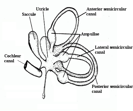
Some components of the vestibular system
Ampullae: a flask-shaped dilatation of the expanded ends of tubular structures referred to as semicircular canals. Each ampulla contains a crista, which is a tranversely oriented ridge of tissue. The upper surface of a crista contain ciliated sensory hair cells embedded in a gelatinous material called the cupula. With movements of the head, the fluid in the ampulla lags behind pushing the cupula slightly, which results in the hairs bending that leads to them stimulate the hair cells, in turning triggering sensory impulses in the vestibular nerve.
Cochlear canal: a spiral tube of the bony labyrinth, which makes about 2.5 turns in human compared, for example, to 1.5 turns in the mouse and 4.5 turns in the guinea pig (an animal famous for being able to right itself to land on all fours when dropped from a height). There are three canals, two of them fluid filled, and the third contains the organ of Corti that detects pressure impulses traveling along the auditory (or acoustic) nerve to auditory cortex. The two fluid-filled ones are the vestibular canal (shown in figure) and the tympanic canal.
Saccule: sensitive to linear translations of head movements in the saggital plane, thus up and down movements as, for example, when moving on an escalator. When the hair cells in a saccule are stimulated, by small stones and a viscous fluid (as for the utricle), a neural impulse is released that travels along the vestibular part of the eighth cranial nerve to the vestibular nuclei in the brain stem. The saccules together with the utricles form the otoliths.
Semicircular canals: involved with the maintenance of the body’s balance and the detection of angular or rotational acceleration of the head. The three canals are located at right angles to each other. While each canal is maximally sensitive to rotations in its own plane, they are organized into functional pairs such that both members of the pair lie in the same plane. Consequently, any rotation in that plane is excitatory to one of the members of that pair, and inhibitory to the other. The ends of the canal connect with the utricle that in turn connects with the saccule. They are structurally developed by about 8 weeks after conception, but it is still a matter of debate as to whether they, and the vestibular system in general, are functional before birth.
Untricle: larger than the saccule, it senses motion in the transverse or horizontal plane, and thus forward-backward, left-right movements and combinations of the two directions. Together with the saccules and semicircular canals, they constitute the vestibular (or membraneous) labyrinths.
See Acceleration, Auditory (or acoustic) nerve, Brain stem, Cochlea, Cranial nerves, Ectoderm, Ganglia, Moro response, Nystagmus, Otoliths, Postural control, Proprioception, Vestibular nuclei, Vestibulo-ocular reflex (VOR), Vestibulospinal tract