Skip to content
The six-layered, 1.4 to 4 mm convoluted sheet or mantle of neural tissue (unmyelinated neurons or tray matter), supported by deep white matter and separated by a prominent, longitudinal central fissure dividing it into in two hemispheres, that dominates the outside view of the human brain and forming the cerebrum. In fact, while it makes up 80% of the brain (but holds only a fifth of its neurons) over 60% of the pallium is ‘buried’ from view. Originally identified by Brodmann in 1909, the six layers or lamina are shown in the figure below. From the most superior (outside) to most inferior (inside) they are: the molecular (I) layer (containing neuroglial cells and receiving nerve fibers from deeper areas), the external (II) granular layer (densely packed with small granular, stellate and pyramidal cells), the external, or medial pyramidal, (III) layer (with pyramidal cells arranged in rows), the internal granular (IV) layer (thinner, but has same cells as the external granular layer, especially small closely packed stellate cells), and which is the primary region of input to the cortex from the periphery, especially the thalamus), the ganglionic (V) layer (a mixture of small granular cells, large pyramidal cells that have output to the brainstem and spinal cord, and cells of some association fibers, which form the bands of Baillarger and Bechterew in the prosencephalon), the fusiform, or multiform, (VI) layer (lots of different neuron types of neurons whose dendrites ascend to the internal granular layer, and whose axons enter the white matter in projecting to the thalamus, but whose function is not clearly understood).
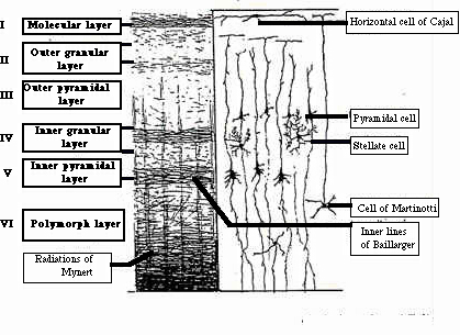
The six layers or lamina of the cerebral cortex after Brodmann
Based on gross morphology, the cerebral cortex is divided into four lobes (each sometimes being referred to as a cortex): frontal lobes, parietal lobes, temporal lobe and occipital lobe (see figure below).
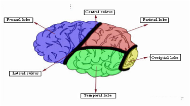
The four lobes of the cerebral cortex. The central sulcus divides the frontal lobe from the other three lobes, and the lateral sulcus the parietal lobe from the temporal lobe. Figure reproduced by kind permission of Patrick McCaffery, California State University, Chico.
Sometimes the limbic system or lobe is treated as a fifth lobe in some classification. Based on the work of 19th and early 20th century histologists, a number of architectonic areas have been identified in the cortex that vary from one of these pioneers to another to the other (differences among the classifications being probably due to normal variations among the brains they analysed), examples of which are as follows:
• Korbinian Brodmann (1868-1918): 49 areas
• Cacile Vogt (1875-1962) & Oskar Vogt (1870-1959): >200 areas
• Constantin von Economo (1876-1931): 109 areas
• A. Walter Campbell (1868-1937): 20 areas
Brodmann’s areal classification or map of ‘postcodes’ tends to be the mostly widely used, which he arrived at sequentially (1 to 49) by electrical probing the cortices of epileptic patients during surgery and noting the behavioral events that resulted form the stimulation (see figure below)
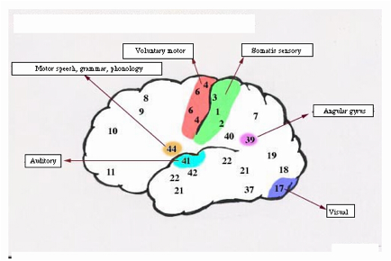
Some of Brodmann’s cortical areas. Figure reproduced by kind permission of Patrick McCafferty, California State University, Chico.
Most of the white matter of the cerebral cortex is made up cortico-cortical connections that are far in excess of the cortico-thalamic and thalamo-cortical connections to the brain stem and spinal cord. Adjacent gyri are connected by U-shaped fibers that run through the white matter. The more distant these connections, the more deeper they are located in the white matter. In the adult human, the total surface area of the cerebral cortex is about 2500 cm², with a thickness varying between 1.5 to 4.5 mm. In the mammalian cortex, the convolutions arise from gyri and grooves referred to as sulci. These allow more surface area for a greater number of neurons while keeping the volume of the brain compact enough to fit inside the cranium. A more literary term than isocortex or neocortex, which refer to the same structure.
See Aphasia, Basal ganglia (anatomy), Basla ganglia (functions), Brain (or encephalon), Brain damage studies, Cerebral cortex (functions), Cortical column, Corpus callosum, Cortical lobes, Equipotentiality, Gray matter, Gyrus, Hormones, Isocortex, Limbic cortices, Limbic system, Locus coeruleus (or ceruleus), Medulla oblongata, Neocortex, Neurogenesis, Neuron, Occiptal cortex (or lobe), Parietal cortex, Prefrontal cortex (PFC), Primary visual cortex (V1), Prosencephalon, Secondary visual cortex (V2), Sulcus, Thalamus, White matter