Growth cone

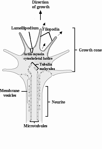
The actively growing, most distal, expanded part of a neurite or outgrowing axon extended from a neuron, and instrumental in target location.  It moves in an ameboid-like fashion, with extensions called filopodia that grow (out) and retract (in) rapidly from the cone due to rapid polymerization and depolymerixation of internal actin filaments, respectively (see figure below for more structural details).  They sense their microenvironment consisting of an array of molecular signals by using guidance receptors (e.g., cadherins, integrins, netrins) on the membrane surface, and move along the extracellular matrix so that the growth cone advances.  First identified by Ramon y Cajal (1832-1934) in 1892, which he considered to be his most prominent discovery. 

Details of a growth cone located at the end of a neurite. The cone is the recipient of membrane vesicles, molecules and proteins transported from the inside of the\growing neurite. Movement of the cone is mediated by the cytoskeletal lattice (found in the lamellipodium between the filopdia) that contains the so-called motor proteins actin and myosin. With extension of the neurite behind the moving cone, the microtubules (forming the backbone of the neurite) are derived from tubulin molecules.  

See Actin, Axon pathway selection, Cell adhesion molecules (CAMs), Extracellular matrix, Fibroblast growth factor (FGF), Filopodia, Lamellipodia, Microtubles, Myosin, Neural cell adhesion molecule (N-CAM), Neurite, Polarizer, Polymerization, Tubulin, Unc-5