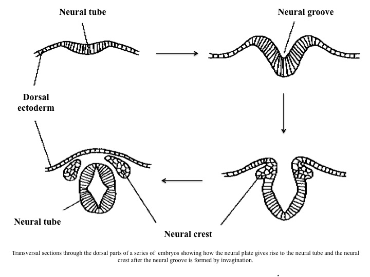
The embryonic precursor or the neural tube containing neural cells in early stages of differentiation, it is one of the rudiments of the central nervous system, which forms from a thickened plate of ectoderm that rolls up around its long axis to form a hollow tubular structure (i.e., the neural tube) extending from the rostral to caudal end of the embryo. Ultimately it will differentiate into the brain and spinal cord. A briefoverview of the main steps in the formation of the neural tube is as follows:
* a dorsal midline strip of primitive ectoderm, the neural plate, rolls itself into a tube. The edges of the plate stick together, closing the tube. Closure begins in the region where the midbrain will later form, and progresses both anterior and posterior, ultimately closing both ends of the tube. Failure to close the neural tube caudally eventuates anencephaly, and rostrally in spina bifida. Then, the next involves;
* the resulting neural tube losing contact with the surface of the embryo and becomes completely surrounded by mesenchyme. The neural tube will become the central nervous system. At the same time, ectoderm immediately lateral to the future neural tube loses contact with the surface, and sinks into the mesenchyme, becoming the neural crests. This tissue generates most of the peripheral nervous system, and also gives rise to the pigment cells of the body, the adrenal medulla, and other non-nervous tissues.
In human embryo, the neural tube closes in week 4 of gestation, and is accompanied by other events crucial to brain development: three primary brain vesicles become evident as does a cephalic flexure, and motor neurons are ‘born’ and migrate away from ventricular surface of spinal cord. Migration of the neural crests haas also commenced, and soon sensory ganglion cells will come into being. The developing relationships among the neural groove, neural crest and neural tube are shown in the figure below:
