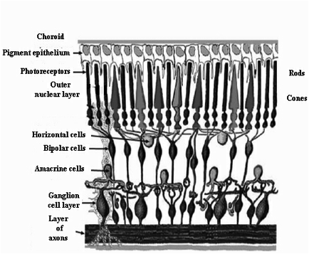
A thin, light-sensitive multilayered pink membrane, the thickness of an onion skin and the size of a postage stamp, lining the inner posterior chamber of the eyeball. The retina itself is lined with a black pigment (melanin) like the inside of a camera is black so as lessen the amount of reflection. This mesh-like tissue contains cones and rods, and is connected by the optic nerve to the primary visual cortex via the lateral geniculate nucleus (see figure below depicting the retina and related structures in the eyeball). It has four major layers: 1. the nuclear layer, further divided into the out nuclear layer in which the ‚aaroots‚aa of the cones and rods are embedded, the outer perform layer containing horizontal and bipolar cells, the inner nuclear layer where amacrine cells reside, the inner perform layer with dendrites from the ganglion cells and the ganglion cell layer, 2. below the ganglion cell layer is that of axons from these cells, 3. above the outer nuclear layer is the photoreceptor layer where the cones and rods reside, 4. on top of this layer is that of the pigmented retinal epithelium, and 5. finally, there is the choroids coat that forms the iris, and which is composed of connective tissue, fibroblasts and a well vascularized layer, the chorio capillaries (see figure below for a representation of these layers). In the ganglion cell layer, there is a small oval area about 2mm in diameter. These cells in this area grow backward to the optic nerve where there are no photoreceptors, and therefore create an optic disc or a ‚aablind spot‚aa (see figure below for a demonstration of the blind spot). Vertebrate retinas arise as outpushings of the brain, and light entering the optic nerve has to traverse through the different neuronal layers before reaching the photoreceptors (an arrangement termed an inverted retina). As mentioned, axons from the ganglion cells then have to travel back through the layers to enter the optic nerve, and it is this that actually creates the ‘blind spot’. Developmentally, the retina appears first as a optic vesicle during the embryonic period due to cell nuclei migrating to the inner surface of the retina. Further development is characterised by the formation of more layers emanating from cell division and subsequent cell migration, and the establishment of multiple complex intercellular connections. An important feature of the retina is that it develops in an inside to outside pattern, with the ganglion cells being formed first and the photoreceptors becoming mature last. By 5 months of gestation, most of the cardinal connections of the retina have been established, and at about this time that the eyelids close to open again during the third trimester of pregnancy. By about 7 months gestation, the eye becomes sensitive to light as demonstrated in preterm infants of this age.