The part of the cerebral cortex, including the primary motor cortex and adjacent areas of the frontal lobe, that generates commands for movement, connected directly to the motoneurons of the spinal cord as well as indirectly to other so-called motor structures such as the basal ganglia and the cerebellum via the thalamus (see figure below). It was Gustav Th. Fritsch (1836-1927) and Eduard Hitzig (1838-1907) in 1870 who together established the motor cortex as fundamental to motor control, which they reported as a paper entitled ‘On the electrical excitability of the cerebrum‘. They did so by applying an electrical current to the precentral gyrus of a dog, and found they could elicit specific movements depending on the location of the stimulus. These connections (or hodology) are schematically represented in the figure below. There is a debate about how the motor cortex ‘codes’ movements. Evidence from non-human primates suggests that populations of cortical neurons code the direction of movement (the ‘population vector hypothesis‘), but this is open to another interpretation, namely, that coding is for the target. Another suggestion is that populations of neurons code for force of movement. While these two suggestions appear to be competitors, it may be the case that they are not mutually exclusive.
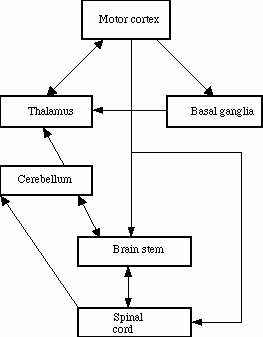
Hodology of the motor system: the basic constituents. The motor system consists of the motor cortex (Brodmann* area 4), forebrain (basal ganglia; cerebellum), midbrain (highest section of brain stem), hindbrain (brain stem; cerebellum), spinal cord (motoneurons), and striated muscles. Not shown are the premotor cortex (Brodmann area 6), supplementary motor area and frontal eye fields (Brodmann area 8), all of which can be considered to be part of the motor system. Note: 1. motor cortex (Brodmann area 4) has direct connections with the spinal cord (decussating corticospinal tracts) and indirect connections (via the brain stem), 2. cerebellum has no direct input from the motor cortex, only one output neuron (viz., Purkinje cells) to the thalamus and brain stem, reciprocal (efferent and afferent) connections with the brain stem and input from the spinal cord, 3. basal ganglia form an integral of the corticothalamic loop and have no input from the spinal cord, and 4. output from the spinal cord goes to innervate the motoneurons of the extrafusal fibers of striated muscles (produce muscle tension during contraction) and the motoneurons to the intrafusal fibers (signal information about the degree of muscle stretch).
*Korbinian Brodmann (1868-1918) who identified distinct areas in the cerebral cortex based on their differences in their cytoarchitecture (i.e., their histology and the arrangement of their six cortical layers).
See Basal ganglia (anatomy), Basal ganglia (functions), Brain stem, Cerebellum (anatomy), Cerebellum and basal ganglia, Cortical column, Corticobulbar tract (CBT), Corticospinal tract (CST), Direct corticomotoneuronal connections (or tracts), Extrafusal muscle fibers, Extrapyramidal system, Frontal eye fields (FEF), Histology, Hodology, Interneurons, Intrafusal muscle fibers, Mirror neurons, Motoneuron, Muscle fiber, Palmar grasp, Premotor cortex, Primary motor cortex, Spinal cord, Striated (or striped or voluntary) muscle, Supplementary motor area (SMA), Thalamus