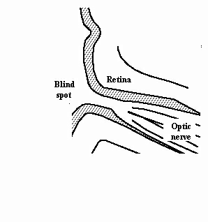
The second pair of 12 cranial nerves, sometimes treated as being part of the central nervous system. Containing 1.2 million ganglion axons, it has far fewer than the number of receptors in the retina, which number above 125 million. This arrangement suggests that quite of large deal of pre-processing takes place in the retina before signals are sent to the brain by means of the optic nerve. The blind spot in the retina corresponds to where the optic nerve leaves the eye (see figure below). It leaves the eye by the optic canal in the direction of the optic chasm. Here, it partially decussates from the temporal visual fields of both eyes to form the optic tracts, and most of its axons terminate in the lateral geniculate nucleus. From this ‘away station’, they travel via the optic radiation to the primary visual cortex in the occipital lobe. Developmentally, the optic nerve makes its first appearance as an out pouching of the diencephalon, and is subsequently sheathed with myelin produced by oligiodendrocytes instead of the usual Schwann cells of the peripheral nervous system.
