First identified by Herophilus of Chaceldon (355-280 BP), the father of anatomy (who also identified the cerebrum and ventricles in humans), it is commonly referred to as the ‘small brain’, and as the ‘head ganglion’ of the proprioceptive system by Charles Sherrington. The cerebellum is a major structure of the metencephalon (hindbrain), lying above and posterior to the medulla oblongata and the pons, that occupies most of the posterior cranial fossa. Consisting of outer layer of gray matter about 2mm thick (the cerebellar cortex) that overlies a central core of white matter, it is connected to the brain stem by three cerebellar pentacles or attachments (see figure below): the inferior cerebellar penducle (or restiform body) containing afferent fibers, the middle cerebellar penducle (or brachium pontis) that also contains afferent fibers, and the superior cerebellar penducle that carries efferent fibers from the deep cerebellar nuclei to the tegementum (dorsal part of the midbrain).
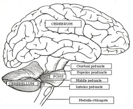
The three cerebellar pentacles. The inferior peduncle receives information from the spiniocerebellar tract, the inferior olivary nucleus, and the reticular formation, and send outputs to the thalamus, reticular formation and vestibular nuclei.The latter convey information about the spatial orientation the head, joints and muscles. The middle penduncle has inputs from the pontine nucleus and relays them to neocerebellum, this being the main pathway from the cerebellum to the cerbellar cortex. The superior peduncle, the major output of the cerebellum, crosses before it gets to the thalamus, and thus influences ipsilateral movements.
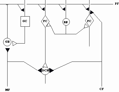
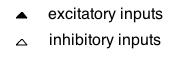
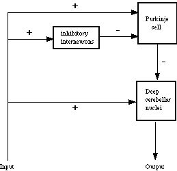
In adult mammals, it makes up about 12% of the total brain mass, and in adult humans it weighs about 142 grams, contains about 70% of the neurons in the brain, and has three-quarters the surface area of the cerebral cortex even though it is much smaller. The latter is achieved through the cerebellar cortex being corrugated by many parallel transverse convolutions or gyri called folia, which are separated from one another by fissures (cf., sulci in the cerebral cortex). Together, the folia and fissures increase the surface of the cerebellar cortex, while at the same time giving it a walnut-like appearance. Three transverse divisions provide one perspective on the gross anatomy of the cerebellum (the other being longitudinal divisions) to reveal three lobes, each having distinct functions. One (the flocclunodular/posterior lobe) is the archicerebellum (or vestibulocerebellum), phylogentically the oldest part of the cerebellum and the first to appear in development, that receives input from the vestibular nerve and the medial and lateral vestibular nuclei. This part of the cerebellum plays a role in the modulation of muscle power and postural control of the antigravity muscles more generally through its influences on the trunk muscles. It also has a role in spatial orientation. Another (the anterior lobe) is the palaeocerebellum (or spinocerebellum) formed by the vermis and anterior parts of the cerebellar hemispheres, which receives inputs from the vestibular system. It too plays an important part in the regulation of muscle power and posture as it receives proprioceptive inputs from the limbs. A third division (the posterior/middle lobe) is the neocerebellum (corticocerebellum or pontocerebellum), phylogentically the newest part of the cerebellum, forming most of the cerebellar hemispheres and part of the vermis and that is the recipient of inputs from the auditory, cutaneous and visual systems. Essential in the coordination of discrete voluntary movements initiated by the cerebral cortex, it is responsible for braking or checking such movements (e.g., reaching). The cortex of the cerebellum is organised into three layers (molecular, Purkinje cell, granular), containing three fibers (climbing, mossy, parallel), and five intrinsic neurons (granule, Golgi type II, Purkinje, parallel, interneurons consisting of basket and stellate cells). The intrinsic circuitry of the cortex involving these neurons, the excitatory and inhibitory inputs to the Purkinje cells, and the inputs and outputs of the cerebellum are illustrated in the following four figures (A-D).
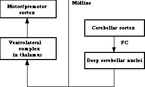
A. Intrinsic circuitry of the cerebellum